LEO1 Polyclonal Antibody
For reference only. Please follow the manual included in your kit for instructions.
Catalog Number
RD89940A

Western blot analysis of extracts of various cell lines using LEO1 Polyclonal Antibody at 1:1000 dilution.
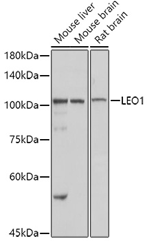
Western blot analysis of extracts of various cell lines using LEO1 Polyclonal Antibody at 1:1000 dilution.
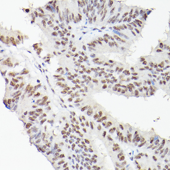
Immunohistochemistry of paraffin-embedded human colon carcinoma using LEO1 Polyclonal Antibody at dilution of 1:100 (40x lens).Perform high pressure antigen retrieval with 10 mM citrate buffer pH 6.0 before commencing with IHC staining protocol.
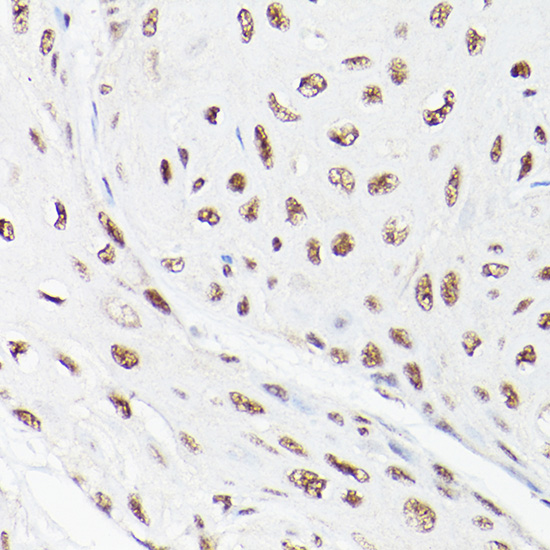
Immunohistochemistry of paraffin-embedded human esophageal cancer using LEO1 Polyclonal Antibody at dilution of 1:100 (40x lens).Perform high pressure antigen retrieval with 10 mM citrate buffer pH 6.0 before commencing with IHC staining protocol.
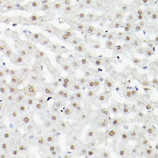
Immunohistochemistry of paraffin-embedded mouse liver using LEO1 Polyclonal Antibody at dilution of 1:100 (40x lens).Perform high pressure antigen retrieval with 10 mM citrate buffer pH 6.0 before commencing with IHC staining protocol.
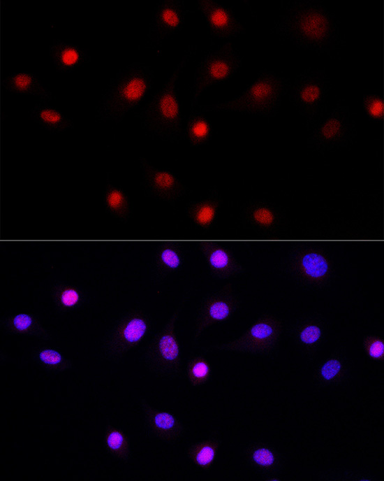
Immunofluorescence analysis of NIH/3T3 cells using LEO1 Polyclonal Antibody at dilution of 1:50 (40x lens). Blue: DAPI for nuclear staining.
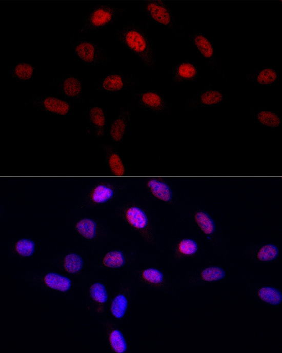
Immunofluorescence analysis of U2OS cells using LEO1 Polyclonal Antibody at dilution of 1:50 (40x lens). Blue: DAPI for nuclear staining.
Product Name
LEO1 Polyclonal Antibody
Catalog Number
RD89940A
Clonality
Polyclonal
Purification Method
Affinity purification
Isotype
IgG
Host
Rabbit
Background
LEO1, parafibromin (CDC73; MIM 607393), CTR9 (MIM 609366), and PAF1 (MIM 610506) form the PAF protein complex that associates with the RNA polymerase II subunit POLR2A (MIM 180660) and with a histone methyltransferase complex (Rozenblatt-Rosen et al., 2005 [PubMed 15632063]).[supplied by OMIM, Mar 2008]
Immunogen Information
Immunogen
Recombinant fusion protein of human LEO1
Gene ID
123169
Swissprot
Q8WVC0
Synonyms
LEO1RDL
Calculated MW
36kDa
Observed MW
45-50kDa
Applications
Reactivity
Human,Mouse,Rat
Tested Applications
WB,IHC,IF
Conjugation
Unconjugated
Dilution
WB 1:500-1:1000,IHC 1:50-1:200,IF 1:50-1:200
Concentration
1mg/mL
Storage Buffer
PBS with 0.01% thiomersal,50% glycerol,pH7.3.
Storage Instructions
Store at -20°C Valid for 12 months. Avoid freeze / thaw cycles.