PDIA4 Polyclonal Antibody
For reference only. Please follow the manual included in your kit for instructions.
Catalog Number
RD89618A
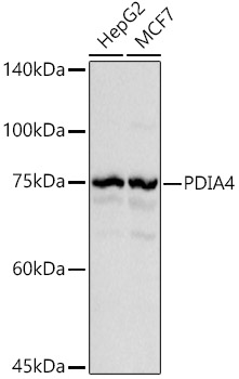
Western blot analysis of extracts of various cell lines using PDIA4 Polyclonal Antibody at 1:1000 dilution.
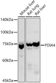
Western blot analysis of extracts of various cell lines using PDIA4 Polyclonal Antibody at 1:1000 dilution.
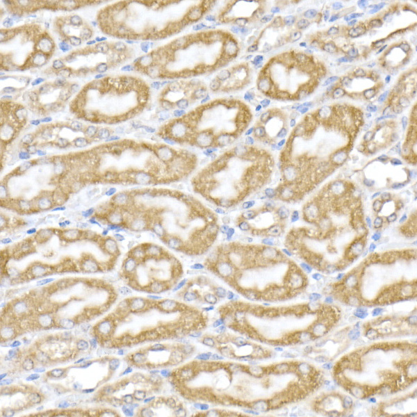
Immunohistochemistry of paraffin-embedded mouse kidney using PDIA4 Polyclonal Antibody at dilution of 1:50 (40x lens).Perform high pressure antigen retrieval with 10 mM citrate buffer pH 6.0 before commencing with IHC staining protocol.
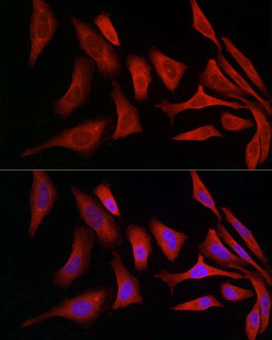
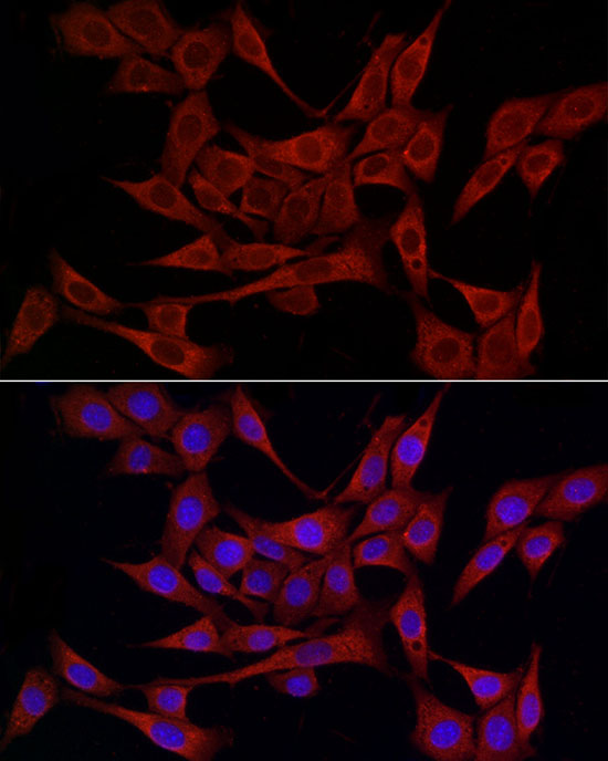
Immunofluorescence analysis of HeLa cells using PDIA4 Polyclonal Antibody at dilution of 1:50 (40x lens). Blue: DAPI for nuclear staining.
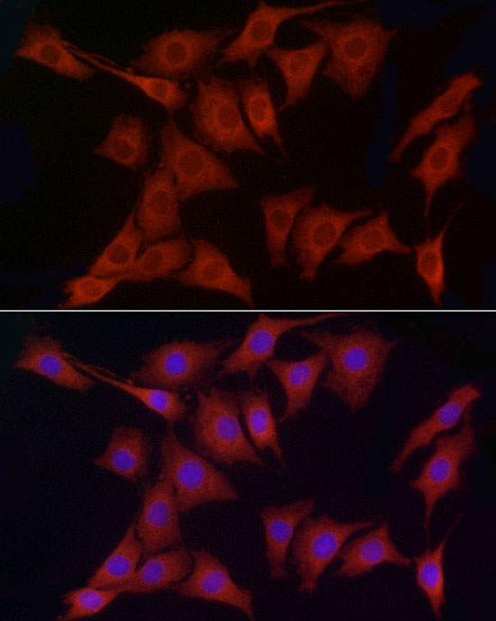
Immunofluorescence analysis of NIH/3T3 cells using PDIA4 Polyclonal Antibody at dilution of 1:50 (40x lens). Blue: DAPI for nuclear staining.
Immunofluorescence analysis of PC-12 cells using PDIA4 Polyclonal Antibody at dilution of 1:50 (40x lens). Blue: DAPI for nuclear staining.
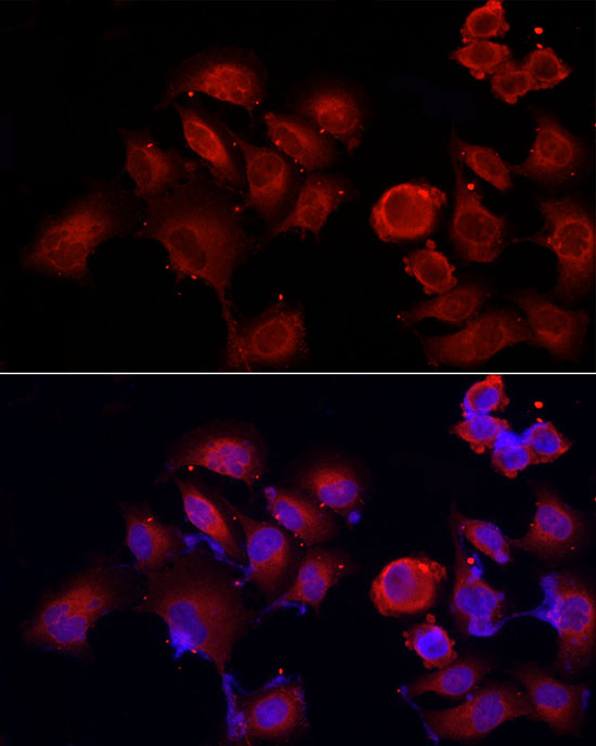
Immunofluorescence analysis of PC-3 cells using PDIA4 Polyclonal Antibody at dilution of 1:50 (40x lens). Blue: DAPI for nuclear staining.
Product Name
PDIA4 Polyclonal Antibody
Catalog Number
RD89618A
Clonality
Polyclonal
Purification Method
Affinity purification
Isotype
IgG
Host
Rabbit
Background
This gene encodes a member of the disulfide isomerase (PDI) family of endoplasmic reticulum (ER) proteins that catalyze protein folding and thiol-disulfide interchange reactions. The encoded protein has an N-terminal ER-signal sequence, three catalytically active thioredoxin (TRX) domains, two TRX-like domains and a C-terminal ER-retention sequence. This protein, when bound to cyclophilin B, enhances the rate of immunoglobulin G intermolecular disulfide bonding and antibody assembly.
Immunogen Information
Immunogen
A synthetic peptide of human PDIA4
Gene ID
9601
Swissprot
P13667
Synonyms
PDIA4ERP70ERP72ERp-72
Calculated MW
52kDa/53kDa
Observed MW
54kDa
Applications
Reactivity
Human,Mouse,Rat
Tested Applications
WB,IHC,IF
Conjugation
Unconjugated
Dilution
WB 1:500-1:2000,IHC 1:50-1:200,IF 1:50-1:200
Concentration
1mg/mL
Storage Buffer
PBS with 0.05% proclin300,50% glycerol,pH7.3.
Storage Instructions
Store at -20°C Valid for 12 months. Avoid freeze / thaw cycles.